Molecular Dynamics

Full GROMACSMolecular Dynamics Pipeline.
Real-Time 3D Visualization.

Complete molecular dynamics simulation from topology generation to MM-PBSA binding energy calculation. Three force fields, automated trajectory analysis, and interactive 3D visualization with 3Dmol.jsinteractive 3D.

End-to-End MD Workflow

From raw PDB structure to binding energy calculation. Every stage is automated, validated, and reproducible.

Stage 01
Topology Generation
Convert PDB to GROMACSmolecular dynamics engine topology. Assign force field parameters, define atom types, and generate position restraints.
Stage 02
Solvation
Place protein in a cubic or dodecahedral box. Fill with TIP3P/SPC water model. Set minimum distance to box edge (1.0-1.2 nm).
Stage 03
Ion Addition
Neutralize system charge with Na+/Cl- counterions. Add physiological salt concentration (150 mM NaCl).
Stage 04
Energy Minimization
Steepest descent minimization to remove steric clashes. Converge to maximum force < 1000 kJ/mol/nm.
Stage 05
NVT Equilibration
100 ps constant volume/temperature equilibration. V-rescale thermostat at 300K. Position restraints on protein heavy atoms.
Stage 06
NPT Equilibration
100 ps constant pressure/temperature equilibration. Parrinello-Rahman barostat at 1 bar. Validate density convergence.
Stage 07
Production MD
10-100 ns production run. 2 fs timestep with LINCS constraints. PME electrostatics. Save coordinates every 10 ps.
Stage 08
Trajectory Analysis
RMSD, RMSF, radius of gyration, hydrogen bond analysis, secondary structure evolution (DSSP), PCA of conformational dynamics.
Stage 09
MM-PBSA Binding Energy
Calculate binding free energy decomposition: van der Waals, electrostatic, polar solvation, non-polar solvation, and entropy contributions.
Stage 10
3D Visualization
Interactive 3Dmol.jsinteractive 3D viewer with hydrogen bonds, binding hotspots, electrostatic surface, and trajectory animation. Exportable as images and movies.

Three Production Force Fields

Choose the force field best suited to your system. Each has been extensively validated for different biomolecular classes.

AMBER99SB-ILDNAll-Atom Protein FF
All-Atom Protein FF
Gold standard for protein simulations. Improved backbone torsion parameters (ILDN). Excellent for protein folding and conformational sampling.
CHARMM27Nucleic Acid FF
Nucleic Acid Compatible
Optimized for protein-nucleic acid systems. CMAP corrections for backbone. Compatible with CHARMM-GUI generated topologies for complex assemblies.
OPLS-AASmall Molecule FF
Small Molecule Optimized
Optimized for organic molecules and drug-like compounds. Excellent Lennard-Jones parameters for protein-ligand binding free energy calculations.

Interactive 3D Analysis

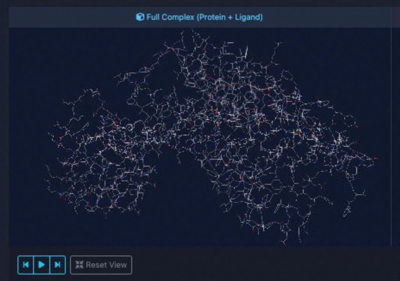
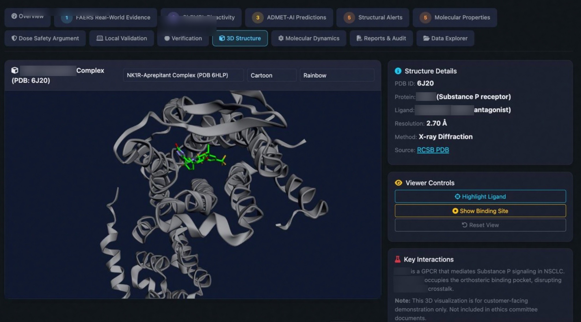
Every MD simulation includes interactive 3Dmol.jsinteractive 3D visualizations showing hydrogen bonds, binding hotspots, and real-time trajectory playback.

3dmol_viewer.html — MD Trajectory Analysis
H-Bonds
14 stable
RMSD
1.8 +/- 0.3 A
Binding dG
-42.3 kJ/mol
Trajectory
50 ns / 5000 frames

MM-PBSA Free Energy Decomposition

Quantitative binding energy calculation with per-residue decomposition. Identify the key interactions driving ligand binding.

Energy Components

The MM-PBSA method decomposes the binding free energy into physically meaningful components, revealing whether binding is driven by electrostatics, van der Waals contacts, or desolvation.

dGbind = dEMM + dGsolv - TdS
vdW
Van der Waals
Lennard-Jones interactions between protein and ligand
Elec
Electrostatic
Coulomb interactions from charged/polar groups
PB
Polar Solvation
Poisson-Boltzmann implicit solvent contribution
SA
Non-Polar Solvation
Solvent-accessible surface area hydrophobic term
-TS
Entropy
Conformational entropy penalty from binding

Ready for Dynamics?

Submit your protein-ligand complex for a full molecular dynamics simulation with binding energy analysis and interactive 3D visualization.